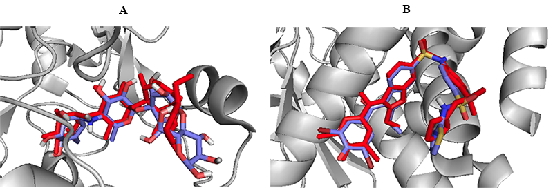
Figure 2. Redocking the co-crystallized ligands on (A) α-glucosidase (B) PTP1B enzymes as a validation step during the docking experiment. The co-crystallized ligands (red) versus docked ligand (blue) are shown in stick representation. A comparable binding orientation was regenerated within same binding pocket by the molecular docking methodology.