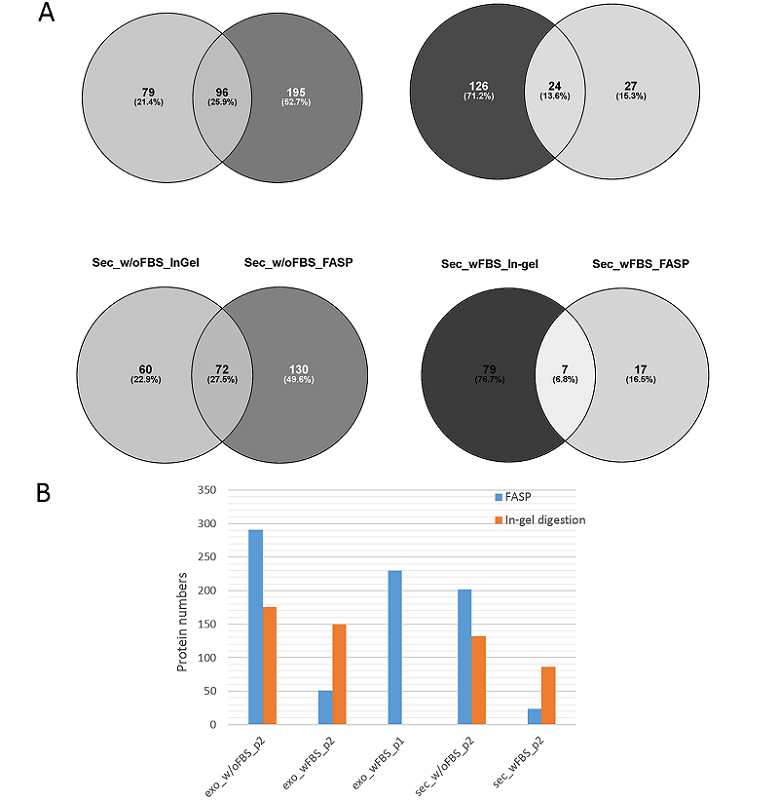
Figure 3. A. Venn diagrams based on the results of mass spectrometric analysis of exosomes (Exo) and secretome (Sec) of Caco-2 colorectal adenocarcinoma cell culture grown in the medium with (wFBS) and without FBS (w/oFBS). Hydrolysis of the samples was performed with in-gel prefractionation (In-gel) and without fractionation on centrifugal filters (FASP). A darker color shows more identifications. B. Total number of proteins identified in exosome (Exo) and secretome (Sec) samples. Two different protocols p1 and p2 were used to isolate exosomes.