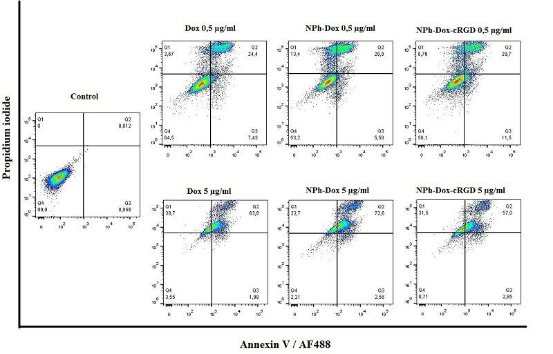

Figure 2. Apoptosis assay in U-87 MG cells culture incubated with free Dox; Dox embedded in phospholipid NPs (NPh-Dox); and Dox embedded in a phospholipid composition with a targeted peptide (NPh-Dox-cRGD). Dox concentrations were 0.5 and 5 µg/ml. Quadrant design: upper left (Q1) – necrosis, cells stained with propidium iodide; top right (Q2) – late apoptosis; bottom right (Q3) – early apoptosis, cells stained with Annexin V; bottom left (Q4) – fluorescence signal at the level of autofluorescence of unstained cells.