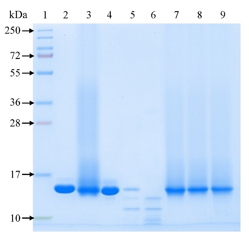
Figure 1. Electrophoregram of the products resulting from trypsinolysis of rhGM-CSF preparations. Electrophoresis in 15% PAAG under non-reducing conditions, Coomassie R-250 staining. Tracks: 1 – protein molecular weight marker (10-250 kDa); 2 – rhGM-CSF, 5 µg; 3 – rhGM-CSF conjugate, 5 µg; 4 – rhGM-CSF + trypsin (100:1); 5 – rhGM-CSF + trypsin (40:1); 6 – rhGM-CSF + trypsin (20:1); 7 – rhGM-CSF conjugate + trypsin (100:1); 8 – rhGM-CSF conjugate + trypsin (40:1); 9 – rhGM-CSF conjugate + trypsin (20:1).