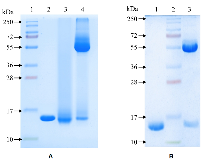
Figure 2. Electrophoregram of dosage forms of rhGM-CSF and its conjugate with polyglucan. Electrophoresis in 15% PAAG under non-reducing conditions, Coomassie R-250 staining. Tracks: A: 1 – rhGM-CSF substance, 10 µg; 2 – rhGM-CSF conjugate, 10 µg; 3 – protein molecular weight marker (10-250 kDa); 4 – dosage form of rhGM-CSF conjugate, 20 µl (3 µg of the target protein). B: 1 – rhGM-CSF substance, 10 µg; 2 – protein molecular weight marker (10-250 kDa); 3 – dosage form of rhGM-CSF, 20 µl (3 µg of the target protein). .