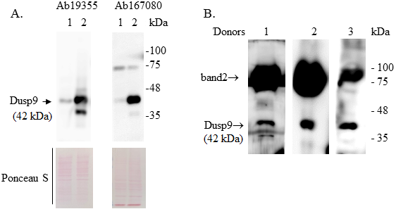
Figure 1. Western blot of proteins extracted from ACHN cells 48 hours after transfection (A) and urine samples from 3 healthy donors (B). A. 1 - ACHN cells transfected with an empty vector and 2 - cells transfected with a vector containing the DUSP9 gene. The positions of the molecular weight marker proteins are shown on the right. Ponceau S staining of membranes was used to control equal protein loading. Ab194355 and Ab167080 – monoclonal and polyclonal rabbit antibodies to DUSP9 (Abcam). B. Proteins of the pellets obtained by centrifugation of 15 ml of urine at 10000 g for 10 min at 20°C were extracted in RIPA buffer. DUSP9 protein was detected using Ab194355.