Age-Related Correlations of Telomere Length of Predator Fish Muscle Tissues with Potentially Different Ageing Mechanisms
Institute of Biomedical Chemistry, 10 Pogodinskaya str., Moscow, 119121 Russia; *e-mail: fireaxe@mail.ru
Keywords: fish aging; pike (Esox lucius); zander (Sander lucioperca); telomere length; muscle tissue physiology
DOI:10.18097/BMCRM00222
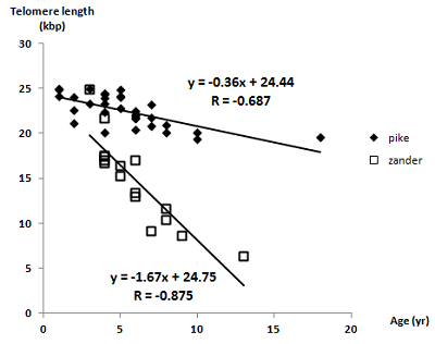
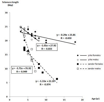
The mechanisms of aging differ and have their own features both mammals, and in different species groups of fish. Telomere length is an indicator of the theoretical number of cell cycles that cells of a particular tissue can go through; therefore, the age-related dynamics of telomere length characterizes changes in the tissue’s ability to regenerate and is necessary to describe the mechanism of tissue aging. In this work, age-related linear regressions of the telomere lengths of muscle tissue of northern pike (Esox lucius) and zander (Sander lucioperca) were empirically obtained for the wide age groups of individuals of both sexes. The identified significant difference in the dependences on their slope values indicates different degrees of decrease in the ability to regenerate muscle tissue with age, which is consistent with the previously discovered physiological characteristics of the muscle tissue of pike. In both fish species studied, telomere length in females decreases with age much more slowly than in males, which is a common feature in the aging mechanisms of most vertebrates.


|
Figure 1.
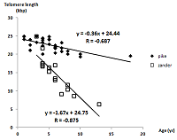
Telomere lengths (in thousands of nucleotide pairs) as a function of age for pike and zander, irrespective of the sex of the fish.
|
FUNDING
The work was performed within the framework of the Program for Basic Research in the Russian Federation for a long-term period (2021-2030) (No. 122030100170-5).
REFERENCES
- Pamplona, R., Jové, M., Gómez, J., Barja, G. (2023) Programmed versus non-programmed evolution of aging. What is the evidence? Exp. Gerontol., 175, 112162. DOI
- Olshansky, S.J. (2018) From lifespan to healthspan. JAMA, 320(13), 1323–1324. DOI
- Reznick, D., Ghalambor, C., Nunney, L. (2002) The evolution of senescence in fish. Mech. Ageing Dev., 123(7), 773–789. DOI
- Bidder, G.P. (1932) Senescence. Br. Med. J., 2(3742), 583–585. DOI
- Lorenzen, K. (2022) Size- and age-dependent natural mortality in fish populations: Biology, models, implications, and a generalized length-inverse mortality paradigm. Fisheries Research., 255, 106454. DOI
- Sauer, D.J., Heidinger, B.J., Kittilson, J.D., Lackmann, A.R, Clark, M.E. (2021) No evidence of physiological declines with age in an extremely long-lived fish. Sci. Rep., 11(1), 9065. DOI
- Finch, C.E. (1990) Longevity, Senescence, and the Genome. Chicago and London: The University of Chicago Press, 938 p.
- Patnaik, B.K., Mahapatro, N., Jena, B.S. (1994) Ageing in fishes. Gerontology, 40(2–4), 113–132. DOI
- Mattern, K.A., Swiggers, S.J., Nigg, A.L., Löwenberg, B., Houtsmuller, A.B., Zijlmans, J.M. (2004) Dynamics of protein binding to telomeres in living cells: Implications for telomere structure and function. Mol. Cell. Biol., 24(12), 5587–5594. DOI
- Rubtsova, M.P., Vasilkova, D.P., Malyavko, A.N., Naraikina, Y.V., Zvereva, M.I., Dontsova, O.A. (2012) Telomere lengthening and other functions of telomerase. Acta Naturae, 4(2), 44–61. DOI
- Forsman, A., Tibblin, P., Berggren, H., Nordahl, O., Koch-Schmidt, P., Larsson, P. (2015) Pike Esox lucius as an emerging model organism for studies in ecology and evolutionary biology: A review. J. Fish Biol., 87(2), 472–479. DOI
- Frost, W.E., Kipling, C. (1959) The determination of the age and growth of pike (Esox lucius L.) from scales and opercular bones. ICES J. Marine Sci., 24(2), 314–341. DOI
- Pravdin, I.F. (1966) Rukovodstvo po izucheniyu ryb. Pishchevaya promyshlennost’, 374 p.
- Steinmetz, B., Müller, R. (1991) An atlas of fish scales and other bony structures used for age determination: Non-salmonid species found in European fresh waters. Samara Publishing, 51 p.
- Cawthon, R.M. (2002) Telomere measurement by quantitative PCR. Nucleic Acids Res., 30(10), e47. DOI
- O'Callaghan, N.J., Fenech, M. (2011) A quantitative PCR method for measuring absolute telomere length. Biological Procedures Online, 13, 3. DOI
- Gil, M.E., Coetzer, T.L. (2004) Real-time quantitative PCR of telomere length. Mol. Biotechnol., 27(2), 169–172. DOI
- Akiyama, M., Yamada, O., Kanda, N., Akita, S., Kawano, T., Ohno, T., Mizoguchi, H., Eto, Y., Anderson, K.C., Yamada, H. (2002) Telomerase overexpression in K562 leukemia cells protects against apoptosis by serum deprivation and double-stranded DNA break inducing agents, but not against DNA synthesis inhibitors. Cancer Lett., 178(2), 187–197. DOI
- Chaddock, R.E. (1925) Principles and Methods of Statistics. Houghton Mifflin, 471 p.
- Aix, E., Gallinat, A., Flores, I. (2018) Telomeres and telomerase in heart regeneration. Differentiation, 100, 26–30. DOI
- Simide, R., Angelier, F., Gaillard, S., Stier, A. (2016) Age and heat stress as determinants of telomere length in a long-lived fish, the Siberian sturgeon. Physiol. Biochem. Zool., 89(5), 441–447. DOI
- Lund, T.C., Glass, T.J., Tolar, J., Blazar, B.R. (2009) Expression of telomerase and telomere length are unaffected by either age or limb regeneration in Danio rerio. PLoS ONE, 4(11), e7688. DOI
- Ocalewicz, K. (2013) Telomeres in fishes. Cytogenetic Genome Res., 141(2–3), 114–125. DOI
- Anchelin, M., Murcia, L., Alcaraz-Pérez, F., García-Navarro, E.M., Cayuela, M.L. (2011) Behaviour of telomere and telomerase during aging and regeneration in zebrafish. PLoS ONE., 6(2), e16955. DOI
- Lopez de Abechuco, E., Hartmann, N., Soto, M., Díez, G. (2016) Assessing the variability of telomere length measures by means of Telomeric Restriction Fragments (TRF) in different tissues of cod Gadus morhua. Gene Reports, 5, 117–125. DOI
- Hatakeyama, H., Nakamura, K.-I., Izumiyama-Shimomura, N., Ishii, A., Tsuchida, S., Takubo, K., Ishikawa, N. (2008) The teleost Oryzias latipes shows telomere shortening with age despite considerable telomerase activity throughout life. Mech. Ageing Dev., 129(9), 550–557. DOI
- Lin, J., Epel, E., Cheon, J., Kroenke, C., Sinclair, E., Bigos, M., Wolkowitz, O., Mellon, S., Blackburn, E. (2010) Analyses and comparisons of telomerase activity and telomere length in human T and B cells: Insights for epidemiology of telomere maintenance. J. Immunol. Methods, 352(1–2), 71–80. DOI
- Conklin, Q.A., Crosswell, A.D., Saron, C.D., Epel, E.S. (2019) Meditation, stress processes, and telomere biology. Curr. Opin. Psychol., 28, 92–101. DOI
- Clough, S.C., Lee-Elliott, I.E., Turnpenny, A.W.H., Holden, S.D.J., Hinks, C. (2004) Swimming speeds in fish: phase 2. Environment Agency, 93 p.
- Mikhailova, M.V., Belyaeva, N.F., Kozlova, N.I., Zolotarev, K.V., Mikhailov, A.N., Berman A.E., Archakov A.I. (2017) Protective action of fish muscle extracts against cellular senescence induced by oxidative stress. Biomeditsinskaya Khimiya, 63(4), 351–355. DOI
- Lapin, A.A., Zelenkov, V.N., Zolotarev, K.V., Mikhailov, A.N., Bodoev, N.V., Mikhaylova, M.V. (2021) Total antioxidant activity of extraction products of muscles and roe of northern pike (Esox lucius). Butlerov Communications С, 1(1), 7. DOI
- Ludlow, A.T., Spangenburg, E.E., Chin, E.R., Cheng, W.-H., Roth, S.M. (2014) Telomeres shorten in response to oxidative stress in mouse skeletal muscle fibers. J. Gerontol. A Biol. Sci. Med. Sci., 69(7), 821–830. DOI
- Dantzer, B., Fletcher, Q.E. (2015) Telomeres shorten more slowly in slow-aging wild animals than in fast-aging ones. Exp. Gerontol., 71, 38–47. DOI
- Adwan Shekhidem, H., Sharvit, L., Leman, E., Manov, I., Roichman, A., Holtze, S., Huffman, D.M., Cohen, H.Y., Bernd Hildebrandt, T., Shams, I., Atzmon, G. (2019) Telomeres and longevity: Acause or an effect? Int. J. Mol. Sci., 20(13), 3233. DOI
- Beirne, C., Delahay, R., Hares, M., Young, A. (2014) Age-related declines and disease-associated variation in immune cell telomere length in a wild mammal. PLoS ONE, 9(9), e108964. DOI
- Leonida, S.R.L., Bennett, N.C., Leitch, A.R., Faulkes, C.G. (2020) Patterns of telomere length with age in African mole-rats: New insights from quantitative fluorescence in situ hybridisation (qFISH). PeerJ, 8, e10498. DOI
- Gardner, M., Bann, D., Wiley, L., Cooper, R., Hardy, R., Nitsch, D., Martin-Ruiz, C., Shiels, P., Sayer, A.A., Barbieri, M., Bekaert, S., Bischoff, C., Brooks-Wilson, A., Chen, W., Cooper, C., Christensen, K., de Meyer, T., Deary, I., Der G., Diez Roux, A., Fitzpatrick, A., Hajat, A., Halaschek-Wiener, J., Harris, S, Hunt, S.C., Jagger, C., Jeon, H.-S., Kaplan, R., Kimura, M., Lansdorp, P., Li, C., Maeda, T., Mangino, M., Nawrot, T.S., Nilsson, P., Nordfjall, K., Paolisso, G., Ren, F., Riabowol, K., Robertson, T., Roos, G., Staessen, J.A., Spector, T., Tang, N., Unryn, B., van der Harst, P., Woo, J., Xing, C., Yadegarfar, M.E., Park, J.Y., Young, N., Kuh, D., von Zglinicki, T., Ben-Shlomo, Y., Halcyon study team (2014) Gender and telomere length: Systematic review and meta-analysis. Exp. Gerontol., 51, 15–27. DOI

