Evaluation of the Collateral Activity of CRISPR/Cas13a-Ribonuclease on the Agilent 2100 Bioanalyzer
Institute of Biomedical Chemistry, 10 Pogodinskaya str., Moscow, 119121 Russia; *e-mail: radkos@yandex.ru
Keywords: CRISPR/Cas13a-ribonuclease; collateral activity; detection; Agilent 2100 bioanalyzer
DOI:10.18097/BMCRM00169
The paper describes an original technique for detecting the collateral activity of CRISPR/Cas 13a ribonuclease based on the assessment of ribosomal RNA degradation. The Agilent 2100 bioanalyzer is used as an analyzing device. This approach is an alternative to existing detection methods and has a number of advantages over them in the case when a quantitative assessment of activity is not required. On the example of the test sample, the optimal concentrations and ratios of the components of the reaction mixture, which are necessary to obtain the most indicative result, were determined. The proposed technique can be used for qualitative assessment of the activity of recombinant ribonuclease Cas13a preparations obtained under different conditions of heterologous protein expression and purification, as well as for testing guide RNAs.
|
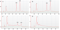
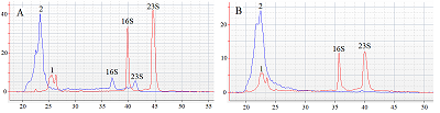
Figure 1.
Electrophoregrams of the E. coli total RNA (panel A) prior and after incubation at 37°C in the presence of RNase A at a concentration of 6·10-9 U/µl for 10, 60 and 120 minutes (panel B, C and D, respectively). 1 is the leading marker; 16S and 23S indicate the peaks of 16S and 23S ribosomal RNA, respectively. The abscissa is time (s); the ordinate is fluorescence (arbitrary units). |
|
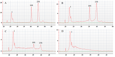
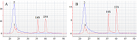
Figure 2.
Electrophoretic analysis of samples containing components of the reaction mixture in various combinations. Panel A - E. coli total RNA; panel B – guide RNA; panel C – a mixture of total RNA and guide RNA; panel D – a mixture of total RNA, guide RNA and Cas13a-nuclease (red electrophoregram – only Cas13a-nuclease; blue – Cas13a-nuclease and guide RNA). The guide RNA and Cas13a concentrations in the samples are 450 and 45 nM, respectively. Samples were incubated for 1 hour at 37°C before analysis. 1 – leading marker; 2 – guide RNA; 16S and 23S indicate the peaks of 16S and 23S ribosomal RNA, respectively. The abscissa is time (s); the ordinate is fluorescence (arbitrary units). |

|
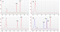
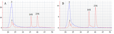
Figure 3.
Electrophoretic analysis of ribosomal RNA fragmentation as a consequence of acquisition of collateral activity by ribonuclease Cas13a. The concentration of Cas13a is 90 nM (panel A) and |
|
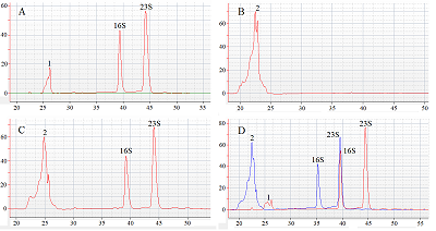
Figure 4.
Effect of Marine RNase Inhibitor on ribosomal RNA fragmentation under the action of Cas13a nuclease. Panel A – in the absence of the inhibitor; panel B – in the presence of the inhibitor |
FUNDING
The study was supported by the Program of Fundamental Research in the Russian Federation for a Long Term of 2021–2030.
REFERENCES
- Gootenberg, J.S., Abudayyeh, O.O., Lee, J.W., Essletzbichler, P., Dy, A.J., Joung, J., Verdine, V., Donghia, N., Daringer, N.M., Freije, C.A., Myhrvold, C., Bhattacharyya, R.P., Livny, J., Regev, A., Koonin, E.V., Hung, D.T., Sabeti, P.C., Collins, J.J., Zhang, F. (2017) Nucleic acid detection with CRISPR-Cas13a/C2c2. Science, 356(6336), 438-442. DOI
- Gootenberg, J.S., Abudayyeh, O.O., Kellner, M.J., Joung, J., Collins, J.J., Zhang, F. (2018) Multiplexed and portable nucleic acid detection platform with Cas13, Cas12a, and Csm6. Science, 360(6387), 439-444. DOI
- Kellner, M.J., Koob, J.G., Gootenberg, J.S., Abudayyeh, O.O., Zhang, F. (2019) SHERLOCK: nucleic acid detection with CRISPR nucleases. Nat. Protoc., 14(10), 2986-3012. DOI
- Myhrvold, C., Freije, C.A., Gootenberg, J.S., Abudayyeh, O.O., Metsky, H.C., Durbin, A.F., Kellner, M.J., Tan, A.L., Paul, L.M., Parham, L.A., Garcia, K.F., Barnes, K.G., Chak, B., Mondini, A., Nogueira, M.L., Isern, S., Michael, S.F., Lorenzana, I., Yozwiak, N.L., MacInnis, B.L., Bosch, I., Gehrke, L., Zhang, F., Sabeti, P.C. (2018) Field-deployable viral diagnostics using CRISPR-Cas13. Science, 360(6387), 444-448. DOI
- Li, Y., Li, S., Wang, J., Liu, G. (2019) CRISPR/Cas Systems towards Next-Generation Biosensing. Trends Biotechnol., 37(7), 730-743. DOI
- Kurbatov, L.K., Radko, P.S., Kravchenko, S.V., Kiseleva, O.I., Durmanov, N.D., Lisitsa, A.V. (2020) Single Stage Purification of CRISPR/Cas13a Nuclease by Metal-Chelating Chromatography Following Heterologous Expression with Preservation of Collateral Ribonuclease Activity. Appl. Biochem. Microbiol., 56(6), 671-677. DOI
- Thormann, W. (2018) Theoretical principles of capillary electromigration methods., in Capillary Electromigration Separation Methods, Poole, C. F., Editor, Elsevier. p. 21-44.